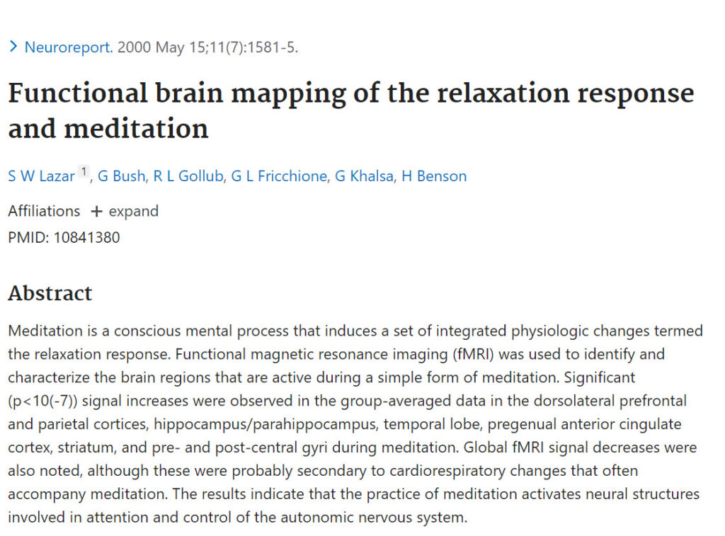
Meditation is a conscious mental process that induces a set of integrated physiologic changes termed the relaxation response. Functional magnetic resonance imaging (fMRI) was used to identify and characterize the brain regions that are active during a simple form of meditation. Significant (p<10(-7)) signal increases were observed in the group-averaged data in the dorsolateral prefrontal and parietal cortices, hippocampus/parahippocampus, temporal lobe, pregenual anterior cingulate cortex, striatum, and pre- and post-central gyri during meditation. Global fMRI signal decreases were also noted, although these were probably secondary to cardiorespiratory changes that often accompany meditation. The results indicate that the practice of meditation activates neural structures involved in attention and control of the autonomic nervous system.
Functional brain mapping of the relaxation response and meditation
Publication
Neuroreport
11(7):1581-5
Abstract
Web and Email Links
Related Listings
Journal
Am. J. Public Health
An experiment conducted at the corporate offices of a manufacturing firm investigated the effects of daily relaxation breaks on five self-reported measures of health, performance, and well-being. For 12 weeks, 126 volunteers filled out daily records and reported bi-weekly for additional measurements. After four weeks of baseline monitoring, they were divided randomly into three groups: Group A was taught a technique for producing the relaxation response; Group B was instructed to sit […]
Journal
Journal of Research and Development in Education
Evaluated self-esteem and locus of control in a group of high school students prior to, during, and following a single academic year. Using a randomized, crossover experimental design, 26 Ss were exposed to either a health curriculum based on elicitation of the relaxation response (RLR) and then a follow-up period, while 24 were assigned to a control health curriculum and then the RLR. Psychological testing was conducted using the Piers-Harris Children's Self Concept Scale and the Now […]
Journal
Biofeedback and Self-regulation
The purpose of this study was to assess the central nervous system effects of the relaxation response (RR) in novice subjects using a controlled, within- subjects design and topographic EEG mapping as the dependent measure. Twenty subjects listened to a RR and control audiotape presented in a counterbalanced order while EEG was recorded from 14 scalp locations. The RR condition produced greater (p < .0164) reductions in frontal EEG beta activity relative to the control condition. N […]

